Tuesday 28 May 2013

Neurodegeneration: Amyloid awakenings : Nature : Nature Publishing Group

Neurodegeneration: Amyloid awakenings : Nature : Nature Publishing Group:

Nature
 
497,
 
S19–S20
 
 
doi:10.1038/497S19a
Published online
 
Sleep disturbances may be an early sign of neurodegenerative diseases — but could sleep deficits cause these conditions in the first place?
Jae-Eun Kang was using a new microdialysis technique to measure how levels of soluble amyloid-β protein in the mouse brain change in response to physiological stress when she noticed something odd. It's thought that the level of soluble amyloid-β correlates with the eventual formation of amyloid-β plaques in the brain, one of the hallmarks of Alzheimer's disease. But Kang found that the protein's concentration seemed to peak during waking hours and fall as the mice slept.
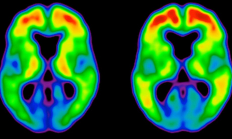
TAMMIE BENZINGER & TYLER BLAZEY, WASHINGTON UNIVERSITY
Patients with preclinical (centre) and clinical (right) Alzheimer's disease show a marked increase in amyloid plaques (red and yellow) in the brain.
Kang, who was a graduate student in neurologist David Holtzman's lab at Washington University in St Louis, Missouri, at the time, then made a further discovery: depriving the mice of sleep led to a dramatic rise in amyloid-β concentration1. “Once we saw that amyloid-β was going up and down with the sleep–wake cycle, the implications began to unfold,” says Holtzman. These findings suggested that sleep disturbances might actually precipitate plaque formation. And if a sleep deficit could increase the concentration of soluble amyloid-β, says Holtzman, then sleep abnormalities in earlier life may predispose people to Alzheimer's.
Holtzman's team went on to show that the natural cycle of waking and sleeping breaks down in mice following plaque formation, but is restored again when antibodies are used to eliminate the plaques2.
This phenomenon is not limited to mice. In people carrying mutations in the presenilin genes, which are strongly linked to the early onset, inherited form of Alzheimer's, Holtzman found that amyloid-β concentration in the cerebrospinal fluid fluctuates to a daily rhythm. He also showed that this cycle breaks down following plaque formation — and before any cognitive symptoms appear2.
So sleep disturbances might be one of the earliest manifestations of Alzheimer's disease, raising the tantalizing possibility of early intervention to help prevent or slow the inevitable march into the cognitive fog.

Early signs

Sleep disturbances might be an early sign of other neurodegenerative conditions as well. Last year, Roxanne Sterniczuk, a neuroscientist at Dalhousie University in Halifax, Canada, looked at data collected from approximately 14,600 people as part of the Survey of Health, Ageing and Retirement in Europe (SHARE), a long-term study of people aged 50 and over across 12 countries.
People who experienced sleep disturbances were more likely to be diagnosed with Alzheimer's within two to four years, according to the SHARE data. In particular, increased daytime sleepiness was the best predictor of the disease. The data also showed that daytime fatigue, a sleep disturbance that leads to exhaustion during the day, predicted those who went on to develop Parkinson's disease.
Parkinson's involves the progressive degeneration of midbrain neurons that produce dopamine — a neurotransmitter involved in regulating the sleep–wake switch. The drugs amphetamine and modafinil, for example, increase wakefulness by increasing dopamine signalling. Conversely, even one night of sleep deprivation reduces the number of dopamine receptors in the striatum, a region of the basal ganglia that is severely affected by the disease. Thus the sleep disturbances associated with Parkinson's may be caused by the degeneration of dopaminergic midbrain neurons.
Sterniczuk plans to analyse the SHARE data again to see if there are subtle differences between the sleep disturbances in presymptomatic Alzheimer's and Parkinson's patients. “If we can characterize the changes before symptoms appear then we can use them as diagnostic markers,” she says. “That might permit earlier treatment.”
The evidence is building for a correlation between disturbed sleep and neurodegenerative diseases, but the next step — discovering whether sleep disturbances are a cause of these conditions — will take considerably more research. In particular, establishing a causal relationship will require longitudinal studies that assess the sleeping patterns of large numbers of people over long periods of time, and link specific types of sleep disorder with the incidence of each disease. But to diagnose the diseases accurately, researchers must look for the tell-tale signs in the brains of study participants.
The Memory and Aging Project, run by David Bennett of Rush University Medical Center in Chicago, Illinois, will allow researchers to do that. The study involves 1,200 elderly people who will undergo annual neurological and psychiatric assessments and have agreed to donate their brains for research. Participants wear wrist monitors that record daily patterns of movement, revealing the times they sleep and wake, and their circadian rhythms. “We'll look at circuitry in the hypothalamus that's involved in sleep and circadian rhythms,” says Clifford Saper, a neurologist at Harvard Medical School in Boston, Massachusetts, who is collaborating on the project. “Presumably, neurodegenerative diseases disrupt sleep and circadian rhythms by damaging this circuitry.”
Testing this hypothesis in humans would be difficult, as it would involve comparing two groups of patients with sleep disorders who are at risk of a neurodegenerative disease, but treating only one group for their sleep problems. “It wouldn't be ethical to withhold treatment from patients, so we'd need to examine how circadian disruption affects neurodegeneration in an Alzheimer's mouse strain,” says Saper. “This would take a couple of years, but it may not mimic what happens in humans.”

Body clock

Sterniczuk is doing just that. In her Alzheimer's model, mice usually develop amyloid-β plaques at about 6 months of age and tau tangles — the other hallmark of Alzheimer's — at 12 months. Before then, their patterns of activity for a particular time of day, such as sleeping and feeding, differ from their non-pathogenic counterparts. The Alzheimer's mice also have fewer neurons in the suprachiasmatic nucleus (SCN), the part of the hypothalamus that regulates the circadian rhythm — a deficiency that makes their circadian clocks go off kilter3.
It's not yet clear whether these changes are associated with altered gene expression in the neurons of the SCN and the raphe nucleus, another part of the brain region that regulates sleep. “I'm trying to find out if changes in the animals' sleep rhythms are associated with altered protein levels in these regulatory regions,” Sterniczuk says. If they are, it raises the possibility of gene therapy. “Targeting these genes may alleviate the sleep and circadian-related symptoms, which may in turn slow the progression of cognitive decline.”
Fruitflies are also yielding clues to the link between neurodegeneration and circadian rhythms. In 2012, researchers at Oregon State University mutated the period gene, which plays a central role in governing the daily circadian rhythm, in fruitflies carrying a mutation in the sniffer gene, which causes neurodegeneration. The flies with both mutations degenerated more quickly, and had shorter lifespans, than flies with either mutation alone, suggesting that disrupted circadian rhythms can accelerate the process of neurodegeneration4.
The findings in mice and flies seem to be directly relevant to humans. In 2005, Jenny Morton, a neurophysiologist at the University of Cambridge, UK, reported that patients with Huntington's disease — a genetic disorder that affects muscle coordination and leads to cognitive decline and psychiatric problems — also have disrupted day–night activity patterns5. Moreover, this disturbance is accompanied by marked falls in the expression of two circadian clock genes in a mouse model of the disease.
Expression of the mouse clock gene mPer2 normally peaks in the morning and dips at night, whereas mBmal1 follows the opposite pattern, being expressed most at night and least in the morning. But in mice carrying genetic mutations associated with Huntington's disease, the expression profiles of both genes break down. In these mice, mPer2 expression is lowest in the afternoon, and the rhythmic expression of mBmal1 ceases altogether. What's more, the expression of both mPer2 and mBmal1 is disrupted in the parts of the brain that first succumb to Huntington's disease: the motor cortex and the striatum. These changes in gene expression are probably a result of disrupted circadian rhythms, rather than a cause of it. “We still don't know whether circadian changes are part of these diseases,” says Morton. “But if they do turn out to exacerbate the neurodegenerative process, then targeting sleep could be of potential therapeutic benefit.”
Genetic variation in clock genes seems to affect people's circadian rhythms, so it may also influence their susceptibility to neurodegeneration. In 2012, for example, Saper's team identified several variations that affect the activity of the human clock gene PER1 and are associated with being either an early riser or a night owl6. “This traces the tendency to be an early riser to a single gene, but we don't know what the implications are for neurodegeneration,” he says.

Broken dreams

Sleeping difficulties and neurodegeneration seem to reinforce one another in a vicious cycle. “Abnormal sleep in mid-life might cause protein aggregation that starts the disease off,” Holtzman says, “and the damage that causes may further disrupt sleep.”
“A lot of early data suggest that modifying sleep could actually delay the onset of disease.”
But could that dynamic be reversed? If disrupted sleep can predispose people to neurodegeneration, then might healthy sleeping patterns help protect the brain against it? Holtzman's group is testing this idea — essentially, whether it is possible to turn the vicious cycle virtuous. “A lot of early data suggest that modifying sleep could actually delay the onset of disease,” he says. “I think that's where the field should be going now, but it's not trivial translating all the animal studies directly into people.”
Holtzman is not alone in thinking this. Earlier this year, Alpar Lazar, who studies sleep and neurodegenerative disease at the University of Cambridge, presented preliminary data showing that bad sleep can precede the onset of Parkinson's disease by many years. His team found that sleep disturbances — particularly fragmented REM sleep — were more severe in patients who are nearer to disease onset than in those who are further away, who in turn had more disturbed sleep than healthy controls.
Huntington's disease is associated with mutations in the huntingtin gene, which contains a short repetitive DNA sequence called a CAG repeat. This repeat sequence is longer in Huntington's patients, leading to the production of misfolded Huntingtin protein. The longer the repeated sequence is, the earlier the disease begins, and the more severe its symptoms. Lazar and colleagues found that longer CAG repeats are also associated with worse sleep disturbances. “Our assumption is that sleep disturbances appear long before disease onset and precipitate disease progression,” says Lazar. So, he says, “intervening to improve sleep may slow down the disease process.” He is conducting a follow-up study to test this hypothesis.
There is already some evidence for this approach. Sleep apnoea, which is characterised by abnormally shallow and interrupted breathing, may almost double the risk of dementia7. Sleep apnoea is treatable, so early intervention could delay the onset not only of neurodegeneration, but also of normal age-related cognitive decline. “Once it is corrected, patients are much brighter and have better memory function,” says Saper, “but it's still not clear whether sleep loss itself increases neurodegeneration.”
Nevertheless, once clinicians wake up to the fact that sleep is so intimately linked to the most common neurodegenerative diseases, they may be in a better position to detect these debilitating neurological conditions at an early stage, and perhaps stop them in their tracks.

Warning signs of, cause OR effect of Alzheimer? | Science | guardian.co.uk

Dreaming of animals and other warning signs of neurodegeneration | Mo Costandi | Science | guardian.co.uk
It is also possible that plaque formation is a consequence of Alzheimer's disease, rather than its cause. According to one new school of thought, it's the soluble form of Aβ protein that is toxic, and the plaques may actually be protecting the brain by capturing these soluble protein particles and preventing them from causing damage. If this turns out to be the case, then blocking plaque formation may actually be harmful. 

Sleep disturbances may be an early warning sign of Alzheimer's and other neurodegenerative diseases

A-beta imagin  
Aβ plaques in the brains of people with preclinical (left) and clinical (right) Alzheimer’s disease. Image: Tammie Benzinger & Tyler Blazey/ Washington University
  The latest issue of Nature contains an Outlook supplement about the health impacts of poor sleep, including a feature I wrote about the link between sleep disturbances and neurodegenerative diseases, called "Amyloid awakenings". The title refers to a process called amyloidosis, by which mutated, abnormally folded proteins aggregate to form insoluble clumps in the brain.

This process is a normal part of ageing, but happens faster in some people than others. Alzheimer's disease, for example, is characterised by insoluble clumps called plaques, which build up in the spaces around neurons in the brain, and neurofibrillary tangles, which accumulate inside the cells. The plaques are made of a mutated protein called amyloid-beta (Aβ), and the tangles of another called Tau. Most other neurodegenerative diseases involve the build-up of misfolded proteins (although each is associated with a different protein or proteins), so amyloidosis does not specifically refer to Aβ aggregation, but is a catch-all term for the process.

The feature grew out of two recent news stories I wrote: the first reported on research presented at the annual meeting of the Society for Neuroscience in New Orleans last October, showing that sleep disturbances may predict Alzheimer's, and the second describes a paper published earlier this year, showing that age-related deterioration of the prefrontal cortex disrupts sleep and impairs memory. Other research published over the past five years or so suggests that sleep disturbances could be an early warning sign of other neurodegenerative conditions, and the article summarises much of this work.

The research shows that people with Alzheimer's, Parkinson's and several other neurodegenerative conditions often experience sleep disturbances many decades before any symptoms appear, and that these disturbances are somehow linked to disruptions of the circadian rhythm. They include common sleeping difficulties such as insomnia, sleep apnoea, and daytime drowsiness, and some slightly more unusual ones. According to one small study published in 2011, for example, the early stages of Parkinson's disease are characterised by alterations in the content of dreams, particularly the presence of animals and increased aggressiveness.

It is still not clear how the sleep disturbances experienced by pre-symptomatic Alzheimer's patients differ from those who will go on to develop one of the other neurodegenerative conditions. Yet, all of the researchers I spoke to seem to agree that sleep disturbances may be the earliest manifestation of these diseases, and that detecting and treating them as early as possible may slow the neurodegenerative process, or even prevent it altogether.

They all agree, too, that the relationship between sleep and neurodegeneration is probably a two-way street. In other words, people with unhealthy sleeping habits earlier on in life may be predisposing themselves to these diseases.

Another new study, published earlier this month, shows that major depressive disorder involves disruption of the activity of hundreds of genes that are involved in regulating the circadian rhythm. Typically, these so-called "clock genes" exhibit regularly fluctuating expression patterns, so that their activity goes up and down with the daily rhythm of the body. As I discuss in my article, the sleep disturbances in patients who go on to develop neurodegenerative diseases are accompanied by a breakdown in the rhythmic expression of clock genes. This new paper is interesting because we now know that depression involves pathological changes similar to those seen in Alzheimer's, including shrinkage of the hippocampus, a part of the brain involved in learning and memory.

One thing I didn't mention in the article, due to space restrictions, is the relationship between protein aggregation and neurodegeneration. In some of these diseases, the misfolded proteins that build up in the brain are highly toxic, and lead directly to neuronal cell death. This is true of the motor neuron diseases and the prion diseases, which include "mad cow disease" and various human forms of it, such as variant Creutzfeldt-Jakob disease (vCJD). For the past few decades, researchers assumed that Aβ plaques cause Alzheimer's disease, and drug companies spent billions researching and developing drugs that block plaque formation or break down plaques that have already formed.

In animal studies, these drugs alleviate the memory impairments associated with Alzheimer's. In humans, however, they don't seem to work, and as a result several large drug companies have halted clinical trials in their late stages. Some researchers are sticking to their guns, arguing that the drugs have to be administered at the earliest stages of the disease to be effective.

It is also possible that plaque formation is a consequence of Alzheimer's disease, rather than its cause. According to one new school of thought, it's the soluble form of Aβ protein that is toxic, and the plaques may actually be protecting the brain by capturing these soluble protein particles and preventing them from causing damage. If this turns out to be the case, then blocking plaque formation may actually be harmful.

How does this come to bear on the link between sleep disturbances and neurodegeneration? In Alzheimer's, plaque formation seems to be closely related to the sleep-wake cycle. One study found that levels of soluble Aβ decrease at night and increase during the day, and are significantly elevated after sleep deprivation. Another showed that the sleep-wake cycle breaks down following plaque formation, but is restored when the plaques are eliminated.

It may mean that the protective mechanism is more active while we sleep than during waking hours, which is in keeping with the emerging view that sleeping well is important for good overall health. More research is needed to clarify exactly how all these factors are related, but this does not bear on the possibility that sleeping difficulties are early diagnostic markers of Alzheimer's and other neurodegenerative diseases.

Sunday 26 May 2013

APOE4 and medium chain triglycerides - Coconut Oil, Ketones and Alzheimer's:

APOE4 and medium chain triglycerides - Coconut Oil, Ketones and Alzheimer's:

Friday, June 19, 2009

APOE4 and medium chain triglycerides

Due to results of studies done by Accera in developing Axona, there is a possible misconception that the medium chain triglycerides will not help people with AD who are APOE4. 
 
Axona has only one of the medium chain triglycerides (C:8) but coconut oil has C:6, C:8, C:10, C:12 (all medium chain triglycerides) and five other fatty acids, including some monounsaturated and polyunsaturated fatty acids, including the essential fatty acid omega-6 (but no omega-3, which is necessary and should be supplemented.) It also happens to have some phytosterol, which is one of the substances that lowers cholesterol and is the basic component in some of the cholesterol lowering agents! It is very possible that each of the fatty acids in coconut oil plays an important and different role in the metabolism of our brains and other organs. 
 
Another possibility is that the Axona folks did not have a huge number of people in their trials and I often wonder if the APOE4 problem was a fluke in their study. 
 
Also, some people in their studies had "mild cognitive impairment," not yet diagnosed with A.D. Dr. Richard Veech of the NIH, an expert in ketones who makes a ketone ester, says that he can think of no biochemical reason why Axona wouldn't work for someone with APOE4. 
 
Steve is APOE4 and responded very well to the medium chain fatty acids. I also read this before trying this with Steve, and therefore expected that MCT oil would not do much for him. I decided to try it anyway in the form of coconut oil for Steve and the rest is history of the happiest kind.

 
posted by Dr. Mary Newport at

Friday 24 May 2013

A Psychosis Caused By Our Immune System | Psychology Today

A Psychosis Caused By Our Immune System | Psychology Today

10% of cases of schizophrenia found to be autoimmune-related in recent study.

brain
Over the past several years, it has become clear that some psychopathology is caused by issues with the immune system, particularly when the immune system attacks our own body, called autoimmune disease. For example, women with post-partum psychosis are more likely than controls to have anti-thyroid antibodies. And folks with schizophrenia and bipolar disorder are more likely to have strange anti-wheat protein antibodies than controls. In the recent, very large CATIE trial,23.% of those with schizophrenia had IgA anti-AGA antibiodies (anti-gliadin) compared to 3.1% of a comparison group, and 5.4% had high levels of tTG antibodies compared to 0.8% of the comparison group. These antibodies are normally tested looking for celiac disease.

No one is sure what these immune reactions mean. But it would be interesting to see how immune modulators might affect psychosis in a clinical trial. In evolutionary medicine, immune and inflammatory modulators could include a dietary intervention, probiotics, or even helminth therapies. All of these could theoretically affect how our immune system reacts to both foreign and domestic (meaning our own) proteins and cell markers. To my knowledge, none of these have been applied to schizophrenia or post-partum psychosis in a clinical trial of any kind.

Earlier this year, a paper came out in the renamed Archives of General Psychiatry (Now JAMA Psychiatry) linking schizophrenia to a set of autoantibodies. The findings in this paper lend more credence to the idea that a subset of schizophrenia may be caused by an immune attack on the brain.  Blood from a group of unmedicated, hospitalized schizophrenics was compared to blood from people admitted with major depressive disorder, borderline personality disorder, and healthy controls.

9.9% of the actuely ill schizophrenics were found to have anti-NMDA receptor antibodies, compared with 2.8% of those with major depressive disorder, 0.4% of controls, and 0 of those with borderline personality disorder. The NMDA receptor (glutamate is the key neurotransmitter at this receptor) is known to be associated with psychotic symptoms. PCP and ketamine are NMDA receptor antagonists that rather famously cause agitation and psychosis.

Find a Therapist

Search for a mental health professional near you.
antibody


Now there is already an illness of anti-NMDA receptors called "NMDA-R encephalitis." At last week's American Psychiatric Association annual meeting, a number of the poster presentations reviewed cases of NMDA-R encephalitis masking as schizophrenia. It typically affects young women with a rare type of ovarian tumor called a teratoma, and presents with psychosis, agitation, memory problems, and seizures. It tends to progress to problems with the autonomic nervous system (which can control breathing, temperature and blood pressure regulation) and cause a catatonic state. It is treated, like many life-threatening autoimmune conditions, with high dose steroids and plasmaphoresis (or plasma exchange, which can clear the blood of the offending autoantibodies).The autoantibodies in the cases of NMDA-R encephalitis are to a different specific protein subunit of the receptor and tend to be in much higher concentrations than the folks with autoantibodies who had acute schizophrenia, so it is not exactly the same disease.  In this trial, however, two of the patients originally diagnosed with schizophrenia were re-diagnosed as NMDA-R encephalitis due to the type of antibodies they had. They also had some intriguing physical symptoms and CNS and blood  inflammatory markers that aren't typically found in schizophrenia.

But it is fascinating and needs to be studied in more populations at greater length. Is there a time coming when 10% of our first break psychosis patients might be getting plasma exchange and steroids? Would they be maintained on autoimmune dietary protocols (if effective for blood titres of antibodies) and relatively benign chronic immune modulators (again, just hypothesizing in an exciting sort of way) such as pig whipworm or killed M vaccae?

As always, more questions than answers, but getting one step closer to the bottom of the pathology of mental illness and brain diseases is always interesting and always gives us hope for new and better therapeutic and preventative options. And what about the healthy control and the patients with major depressive disorder who had anti-NMDA-R antibodies? Are they more likely to have problems with psychosis or psychopathology? We will have to wait and see.
Images from wikimedia commons.
Copyright Emily Deans, M.D.

Tuesday 21 May 2013

Scientists identify molecular trigger for Alzheimer’s disease | University of Cambridge

Scientists identify molecular trigger for Alzheimer’s disease | University of Cambridge

New research establishes nature of malfunction in protein molecules that can lead to onset of dementia.

We have to solve what happens at the molecular level before we can progress and have real impact
Tuomas Knowles
Researchers have pinpointed a catalytic trigger for the onset of Alzheimer’s disease – when the fundamental structure of a protein molecule changes to cause a chain reaction that leads to the death of neurons in the brain.

For the first time, scientists at Cambridge’s Department of Chemistry, led by Dr Tuomas Knowles, Professor Michele Vendruscolo and Professor Chris Dobson working with Professor Sara Linse and colleagues at Lund University in Sweden have been able to map in detail the pathway that generates “aberrant” forms of proteins which are at the root of neurodegenerative conditions such as Alzheimer’s.

They believe the breakthrough is a vital step closer to increased capabilities for earlier diagnosis of neurological disorders such as Alzheimer’s and Parkinson’s, and opens up possibilities for a new generation of targeted drugs, as scientists say they have uncovered the earliest stages of the development of Alzheimer’s that drugs could possibly target.

The study, published today in the Proceedings of the US National Academy of Sciences, is a milestone in the long-term research established in Cambridge by Professor Christopher Dobson and his colleagues, following the realisation by Dobson of the underlying nature of protein ‘misfolding’ and its connection with disease over 15 years ago.

The research is likely to have a central role to play in diagnostic and drug development for dementia-related diseases, which are increasingly prevalent and damaging as populations live longer.

In 2010, the Alzheimer’s Research UK showed that dementia costs the UK economy over £23 billion, more than cancer and heart disease combined. Just last week, PM David Cameron urged scientists and clinicians to work together to “improve treatments and find scientific breakthroughs” to address “one of the biggest social and healthcare challenges we face.”

The neurodegenerative process giving rise to diseases such as Alzheimer’s is triggered when the normal structures of protein molecules within cells become corrupted.

Protein molecules are made in cellular ‘assembly lines’ that join together chemical building blocks called amino acids in an order encoded in our DNA. New proteins emerge as long, thin chains that normally need to be folded into compact and intricate structures to carry out their biological function.

Under some conditions, however, proteins can ‘misfold’ and snag surrounding normal proteins, which then tangle and stick together in clumps which build to masses, frequently millions, of malfunctioning molecules that shape themselves into unwieldy protein tendrils.

The abnormal tendril structures, called ‘amyloid fibrils’, grow outwards around the location where the focal point, or 'nucleation' of these abnormal “species” occurs.

Amyloid fibrils can form the foundations of huge protein deposits – or plaques – long-seen in the brains of Alzheimer’s sufferers, and once believed to be the cause of the disease, before the discovery of ‘toxic oligomers’ by Dobson and others a decade or so ago.

A plaque’s size and density renders it insoluble, and consequently unable to move. Whereas the oligomers, which give rise to Alzheimer's disease, are small enough to spread easily around the brain - killing neurons and interacting harmfully with other molecules - but how they were formed was until now a mystery.

The new work, in large part carried out by researcher Samuel Cohen, shows that once a small but critical level of malfunctioning protein ‘clumps’ have formed, a runaway chain reaction is triggered that multiplies exponentially the number of these protein composites, activating new focal points through ‘nucleation’.

It is this secondary nucleation process that forges juvenile tendrils, initially consisting of clusters that contain just a few protein molecules. Small and highly diffusible, these are the ‘toxic oligomers’ that careen dangerously around the brain cells, killing neurons and ultimately causing loss of memory and other symptoms of dementia.

“There are no disease modifying therapies for Alzheimer’s and dementia at the moment, only limited treatment for symptoms. We have to solve what happens at the molecular level before we can progress and have real impact,” said Dr Tuomas Knowles from Cambridge’s Department of Chemistry, lead author of the study and long-time collaborator of Professor Dobson and Professor Michele Vendruscolo.

“We’ve now established the pathway that shows how the toxic species that cause cell death, the oligomers, are formed. This is the key pathway to detect, target and intervene – the molecular catalyst that underlies the pathology.”

The researchers brought together kinetic experiments with a theoretical framework based on master equations, tools commonly used in other areas of chemistry and physics but had not been exploited to their full potential in the study of protein malfunction before.

The latest research follows hard on the heels of another ground breaking study, published in April of this year again in PNAS, in which the Cambridge group, in Collaboration with Colleagues in London and at MIT, worked out the first atomic structure of one of the damaging amyloid fibril protein tendrils. They say the years spent developing research techniques are really paying off now, and they are starting to solve “some of the key mysteries” of these neurodegenerative diseases.

“We are essentially using a physical and chemical methods to address a biomolecular problem, mapping out the networks of processes and dominant mechanisms to ‘recreate the crime scene’ at the molecular root of Alzheimer’s disease,” explained Knowles.

“Increasingly, using quantitative experimental tools and rigorous theoretical analysis to understand complex biological processes are leading to exciting and game-changing results. With a disease like Alzheimer’s, you have to intervene in a highly specific manner to prevent the formation of the toxic agents. Now we’ve found how the oligomers are created, we know what process we need to turn off.”
Inset image: L-R, Professor Christopher Dobson, Dr Tuomas Knowles and Professor Michele Vendruscolo

For more information, please contact fred.lewsey@admin.cam.ac.uk

Testosterone and Memory - Blog - Testosterone replacement & general men's health articles

Testosterone and Memory - Blog - Testosterone replacement & general men's health articles

Posted by on in Mental Health
 
Memory problems are a very commonly reported symptom of andropause. As with memory loss associated with menopause, it is important to be aware that occasional minor lapses in memory are nothing to worry about.

When loss of memory does affect your ability to function, it may be time to consider getting medical help.

Cognition is the mental process that includes language, calculation, visual-spatial abilities, memory, reasoning, learning, social skills, imagination and attention span. With age, cognitive function may remain stable or decline.

In general, cognitive function that remains stable includes attention span, everyday communication skills, language skills, and simple visual perception. Cognitive function that decline includes selective attention, naming of objects, verbal fluency, complex visualspatial skills and language analysis. Moreover, age-related memory changes vary depending on the type of memory.

The acquisition and early retrieval of recently acquired information is diminished; long-term memory retention does not seem to change with increasing age.

MEMORY LOSS OCCURS DURING ANDROPAUSE

This may be secondary to the effects of declining hormones. With age, the levels of hormones may remain the same, elevated or reduced.

THE CIRCULATING LEVELS OF HORMONES THAT DECLINE IN AGING MALES ARE:
  • TESTOSTERONE: TOTAL AND FREE TESTOSTERONE
  • DEHYDROEPIANDROSTERONE (DHEA) AND DEHYDROEPIANDROSTERONE SULFATE (DHEAS)
  • GROWTH HORMONE AND INSULIN-LIKE GROWTH FACTOR-1 (IGF-1)
  • OTHER HORMONES INCLUDING TRIIODOTHYRONINE, RENIN AND ALDOSTERONE

The effect of hormones on cognition in the older adults has been investigated for several years. Hormone effects in elderly women's cognitive function have been researched in depth.

There has been abundant data on the benefits of estrogen replacement on cognition and mood.
The effect on cognition by estrogen should be considered as longterm and modifying factors and not the cause of Alzheimer's disease. Relatively speaking, there is paucity about the link between the cognitive change and hormones in the older men. The improvement of cognition may assist older adults to remain independent and may contribute to prolong the time to institutionalization.

THE MANIFESTATIONS OF MALE MEMORY LOSS ARE VARIED

Many andropausal men experience difficulty remembering how complete familiar tasks. For instance: forgetting why you went to a store; forgetting what you are trying to say in mid-sentence; or forgetting the reason you went to talk with a co-worker.

While it is clear that genetics play a part in male memory loss, they are usually not the entire picture. Memory loss in andropausal men is not simply an inevitable part of aging, but often a symptom of hormonal imbalance.

MEDICAL TREATMENTS FOR MALE MEMORY LOSS

Lapses in memory and foggy thinking are often symptoms of andropausal memory loss associated with hormonal imbalances.

Andropausal memory loss can be very difficult to cope with, negatively impacting every aspect of your life. It is important to remember that you are not losing your mind: your hormones are simply taking you for a bad ride.

Memory loss in andropausal men that is related to hormonal imbalances can often be medically treated using bioidentical hormone therapy.

Hormone therapy for memory loss in men can contribute to the recovery of normal memory. Further, Andro Medical Group programs are custom tailored with nutrition and supplements plans that support greater overall male health as well as clear thinking.

YOU DON'T HAVE TO ACCEPT MEMORY LOSS AS A PART OF AGING